Digitalisation in histology practicals
 Digital histology
-
©DSimage/KI generiert - stock.adobe.com
Digital histology
-
©DSimage/KI generiert - stock.adobe.com
To recognise innovative approaches to improving the quality of teaching, the Medical University of Graz awards a special grant totalling 50,000 euros each year. One of the award-winning projects is ‘Digitalisation in histology practicals – improving interactive teaching methods and digital support for study groups’.
Knowledge transfer in the pre-clinical sector
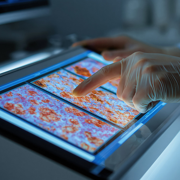
At universities, the use of digital tools for knowledge transfer is becoming increasingly important, particularly as students will also be confronted with digital applications more frequently in their future working environments. To ensure the best possible knowledge transfer and optimal preparation for future postgraduate processes of knowledge acquisition, the Chair of Cell Biology, Histology and Embryology is consistently pursuing its path of digitalisation, which began with the HRSM project ‘Digitalisation of Teaching’. Students now learn, during their histology and embryology practical sessions, not only how to use the analogue microscope and ‘transparent’ tool for structural analysis, as well as how to use digital microscopy via a screen mounted directly next to the microscope, on which they can view the histological sections in high resolution via touchscreen and discuss them together. With this digital component of histology teaching, the division now has the opportunity to display additional rare stains for illustrating different tissue structures, specific stains to illustrate particular cell types and histophysiological relationships within the tissue, as well as electron microscope images that can reveal the cell biology of the cells.
Interactive learning environment
To encourage interaction and discussion amongst students even outside histology lessons, a large touchscreen monitor connected to the histology database has been installed outside the microscopy room. This offers students the opportunity to explore histological structures individually or in small groups outside of practical sessions, and to deepen their understanding of the subject matter. Initial experiences with this additional digital resource clearly show that supplementing the analogue world with the digital one is the right way to convey structural and histophysiological knowledge even more effectively. This is also evident from student feedback, which ranges from increased enthusiasm for histology and praise for the high standard of the histology track to comments that the histology component was the best compulsory track to date. However, direct communication between students and lecturers is crucial to this interactive and learning-oriented environment. Therefore, the digitisation of histology practicals is not about virtualising the content, but about making additional use of digital tools to enhance direct, face-to-face communication.