New standardised framework for tissue mapping accelerates AI-assisted pathology research
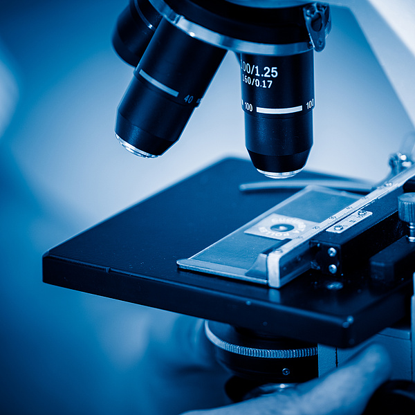 Arbeiten mit dem Mikroskop
-
© alafoto - stock.adobe.com
Arbeiten mit dem Mikroskop
-
© alafoto - stock.adobe.com
As one of the key outcomes of the international EU-funded project ONCOSCREEN, a team of researchers from Medical universities and institutions in Austria and Czech Republic has introduced a framework to standardize metadata for Whole Slide Images (WSIs), significantly advancing the efficiency and interoperability of artificial intelligence (AI) applications in pathology and cancer research. The framework has recently been published in Artificial Intelligence in Medicine.
From slides to maps
Whole Slide Images are high-resolution digital scans of complete glass slides containing biological specimens such as tissue sections or cell samples. These images, captured at multiple magnifications, are digitally viewable, analyzable, and shareable. WSIs have become foundational to AI algorithm development and are widely used in pathology for disease diagnosis and oncology for cancer research. Beyond these areas, they are increasingly applied in neurology, veterinary medicine, hematology, microbiology, dermatology, pharmacology, toxicology, immunology, and forensic science.
Despite their growing importance, a major challenge has limited their full potential: the absence of a standardized system to describe WSI content. Currently, assembling cohorts for AI training and validation requires manual inspection of images to determine their morphological content — an approach that is impractical for modern archives containing millions of images.
To address this gap, the research team proposes a general framework for generating structured 2D index maps, or “tissue maps” that describe the morphological composition of WSIs using a common syntax and semantic structure. The framework is designed to ensure interoperability between WSI catalogs and archives.
The proposed tissue maps are organized into three hierarchical layers:
- Source Layer – Identifies the biological origin of the specimen
- Tissue Type Layer – Classifies the anatomical or histological tissue category
- Pathological Alteration Layer – Annotates disease-related or abnormal morphological features
Making data AI-ready
Each layer assigns image segments to defined classes, producing AI-ready metadata that can be automatically generated and systematically integrated into digital archives.
The researchers demonstrated the effectiveness of the framework by applying AI-based metadata extraction to generate tissue maps from WSIs and incorporating them into an existing WSI archive. The integration substantially enhanced search and filtering capabilities, enabling faster identification of relevant cases and more precise dataset assembly.
“We turn massive digital tissue scans into searchable data. By describing the content of these images with our tissue maps, we help researchers quickly build high-quality datasets required to train next-generation AI tools for healthcare”, Gernot Fiala explains
By standardizing WSI metadata and enabling automated tissue map generation, this framework facilitates the accelerated creation of high-quality, balanced, and targeted datasets for AI training and validation. The approach is expected to improve reproducibility, interoperability, and scalability across digital pathology platforms and research institutions.
This development represents an important step toward fully leveraging large-scale WSI repositories for AI-driven diagnostics and cancer research, ultimately supporting more efficient scientific discovery and improved patient outcomes.